Friday 28 February 2014

CERVICO-ISTHMIC PREGNANCY


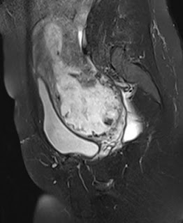
A patient presented in her mid thirties with a first trimester intra-uterine pregnancy having conceived after IVF.  USG showed a fetus with cardiac activity and crown-rump length corresponding to 11 weeks' gestation.  The gestation sac was abnormally low lying with an empty endometrial cavity above it. The cervix was poorly visualized. An MRI was indicated to rule out true cervical pregnancy.

MRI showed a low lying gestation sac with empty endometrial cavity above it. The cervix was shortened with a closed cervical canal and internal os. There was a large low lying placenta surrounding the sac with loss of interface between the placenta and myometrium. The myometrium was thinned out. A diagnosis of cervico-isthmic pregnancy was made with the likelihood of adherent placenta.

CERVICO-ISTHMIC PREGNANCY


INTRODUCTION: A cervico-isthmic pregnancy is an ectopic pregnancy which implants in the isthmus. The isthmus is the region between the cervix and fundus.

It is a rare complication of pregnancy and is often confused with a cervical pregnancy. Distinguishing between these is crucial as a cervical pregnancy is virtually not viable while there are a few reports of cervico-isthmic pregnancies being carried successfully to term. Also, cervical pregnancies are more dangerous and associated with serious risks like haemorrhage, rupture, blood transfusions, hysterectomy etc.

INCIDENCE:
Incidence of cervical pregnancy: 1:1000 to 1:16000.
Cervico-isthmic pregnancies are more common than true cervical pregnancies.

IMAGING FINDINGS:
USG: Typically, ultrasound shows an empty uterine cavity with a low lying pregnancy. It may be confused with true cervical pregnancy. In a true cervical pregnancy, the sac is completely within the cervix.

MRI: shows a  low lying gestation sac with a well preserved and closed cervical canal. It is hence helpful in distinguishing cervical from cervico-isthmic pregnancy.

IMPLICATIONS: Although there are reports of successful cervico-isthmic pregnancies, these pregnacies are also exposed to risks like:
  • Spontaneous abortion/ premature delivery
  • premature rupture of membranes
  • placenta accreta
  • postpartum hemorrhage
  • hysterectomy
  • Uterine rupture if associated with thinned out myometrium
REFERENCES:
  1. Oyelese Y, Elliott T B, Asomani N, Hamm R, Napoli L, Lewis KM: Sonography and Magnetic Resonance Imaging in the Diagnosis of Cervico-Isthmic pregnancy. J Ultrasound Med 2003; 22: 981-983
  2. Strobelt N, Locatelli A, Ratti M, Ghidini A: Cervico-isthmic pregnancy: a case report, critical appraisal of the diagnostic criteria, and reassessment of the outcome. Acta Obstet Gynecol Scand 2001; 80: 586-588.
  3. Jelsema RD, Zuidema L: First trimester diagnosed cervico-isthmic pregnancy resulting in term delivery. Obstet Gynecol 1992; 80: 517-519
  4. Wikipedia


RHOMBENCEPHALOSYNAPSIS

Figure 1: Coronal T2 image showing fused dentate nucleus
Figure 2: Coronal T2 weighted image showing typical transverse folia and sulci with fused cerebellar hemispheres

Figure 3: Axial T2WI showing fusion of the interfoliate sulci across midline along with fusion of cerebellar folia.

Figure 4: Axial T2WI showing "keyhole appearanca" of 4th ventricle and midline fusion of cerebellar white matter. Vermis is not seen.

Figure 5: Sagittal T2WI showIng absent primary fissure. There is also slight rounding of the fastgial recess. Note the corpus callosum in a patient of isolated rhombencephalosynapsis.

The above MR was done in an adult patient presenting with mild gait ataxia. Rhombencephalosynapsis was detected in him as an isolated finding.

  • Rhombencephalosynapsis is a rare  posterior fossa malformation characterized by aplastic/ hypoplastic cerbellar vermis and fusion of cerebellar hemispheres in the midline.
  • It was forst described by Obersteiner in 1914.
  • Pathogenesis is related to disturbed cerebellar development between 28 to 41 days of gestation.
  • Clinical profile varies and is related to the posterior fossa malformation and associated supratentorial anomalies. It might vary from mild truncal ataxia and normal intelligence to cerebral palsy.
  • Isolated rhombencephalosynapsis is less common than rhombencephalosynapsis associated with supratentorial anomalies
  • Most cases are reported in infancy or pediatric age group with only few cases reported in adults.

Diagnosis is usually by MRI which is clearly superior to CT in evaluating these patients.

Distinctive radiological features are:
Posterior fossa:
  • Agenesis / hypoplastic vermis 
  • Absent posterior cerebellar incisura, vallecula
  • agenesis or hypogenesis of anterior vermis ( rostral ) with hypoplastic posterior vermis( caudal  nodulus may form
  • Midline fusion of cerebellar hemispheres to form a single lobed cerebellum
  • Fissures and cerebellar folia are transversely oriented best seen on coronal MRI
  • Superior and middle cerebellar peduncles may be fused along with dentate nuclei giving a characeteristic diamond shaped, posteriorly pointing 4th ventricle
Supratentorial anomalies:
  • Most common supratentorial anomaly is hydrocephalus with aqueductal stenosis
  • Sometimes, there may be fused thalami, fornIces, cerebral peduncles, inferior collicluli with absent septum pellucidum
  • Dysgenetic limbic system
  • cortical malformatins
  • sutural synostosis
  • hypoplastic commissures, hypoplastic anterior visual pathway and agenesis of posterior lobe of pituitary gland
  • Association with septo-optic dysplasia and holoprosencephaly
 References:
  • Mendonca JLF, Natal MRCR, Viana SL et al. Rhombencephalosynapsis: CT and MRI findings. Neurology India;152:118-120, 2004.
  • Weerakkody Y, Gaillard F et al. Rhombencephalosynapsis. Radiopaedia.org.
  • Blaser SI: Rhombencephalosynapsis. Diagnostic Imaging Brain: First Edition. Amirsys: I-1-30, 2005.