Tuesday, 31 December 2013

MRI in Spigelian Hernia


T1 weighted images in axial and sagittal plane, T2 FS images in axial plane of an 80 year old male presenting with palpable swelling in the right ventral abdominal wall shows focal herniation of mesenteric / omental fat and vessels through a defect in the right spigelian aponeurosis ( that of the internal oblique and transversus abdominis muscles) inferior to the umbilicus. The hernial sac is covered externally by an intact aponeurosis of the extrenal oblique muscle. There is no herniation of bowel loops through this defect.
  • Spigelian Hernia is a hernia through a defect in the aponeurosis of internal oblique and transversus abdominis muscles.
  • It is seen seen as a defect in the spigelian aponeurosis  lateral to the rectus muscles, inferior to umbilicus where the sheath is deficient posteriorly.
  • The external oblique aponeurosis is intact with the hernial sac lying in interparietal/ intermuscular plane 
  • It is named after a Belgian surgeon-anatomist by the name of Adriaan van den Spiegel although it was first described by KLINKOSCH
  • Raveenthiran has described the existence of spigelian hernias with cryptorchidism called the Raveenthiran Syndrome
  • It is a rare hernia, approx. 1%( range of 0.2 -2%) of ventral hernias.
  • It is commoner on the right side in 4th-7th decade of life.
  • It is commoner in females with a male to female ratio of approx. 1:1.18.
  • It may be congenital or acquired
  • It may be entirely asymptomatic. Its clinical significance lies in the fact that the hernial opening is small predisposing the bowel loops to strangulation/ obstruction. Hence, it is importatnt to repair it. The repair is usually straightforward and rarely requires a mesh prosthesis.
  • It is difficult to diagnose clinically, especially in obese patients
  • Imaging: USG is often used in conjunction with Valsalva's manouvre in real time to demonstrate the hernia. CT is considered optimal especially with oral contrast. The role of MRI is less described in literature being a more expensive modality. However, with the wider availability of MRI and its better anatomical visualization, it may prove useful in preoperative evaluation as in this patient.
  • Differentials are ventral hernias, umbilical hernia, hernia thorugh laparoscopy port etc.
REFERENCES:
  • Harrison LA, Keeshing CA, Martin NL et al. Abdominal wall hernias: Overview of herniography and correlation with cross-sectional imaging. Radiographics 1995; 15(2):315-22.
  • Mittal T, Kumar V, Khullar R et al: Diagnosis and management of Spigelian Hernia: A review of literature and our experience. J. Minimum Access Surgery 2008; 4(4): 95-98
  • Raveenthiran V. Congenital spigelian hernia with Cryptorchidism: Probably a new syndrome. Hernia 2005; 9(4):378-80
  • Federle MP, Jeffrey, RB, Woodward PJ, Borhani AA. Spigelian Hernia. Diagnostic Imaging Abdomen: Second Edition. Amirsys: II-1, 37,2010


Saturday, 30 November 2013

SIMPLE BONE CYST


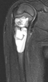
T1 and STIR coronal and sagittal MR images in a 4 year old child showing a fairly well demarcated expansile septate cystic lesion in the proximal metaphyseal region of right humerus. It displays mild heterogeneity but is largely homogenous.

SIMPLE BONE CYST: 
-90% occur in long bones, most common in proximal humerus
-Metaphyseal expansile cystic lesion
-May contain "fallen fragment" which is  a fractured fragment of bone which moves in dependent part with change in patient position. It is usually seen on x-rays and CT.
-May show fluid-fluid level on CT and MRI
-"Rising bubble sign" : Bubble of gas in non-dependent part of lytic lesion suggestes pathologic fracture. It may be seen on CT and MRI.
-FDG active, may hence mimic metastasis on PET-CT

REFERENCE:
Manaster BJ, Roberts CC, Petersilge CA, Moore S, Hanrahan CJ, Crim J.Diagnostic Imaging Musculoskeletal: Non-traumatic disease. Amirsys.2-206, 2010

Focal dehiscnece of lamina papyracea


Above axial CT images show focal dehiscence / absence of lamina papyracea in the right orbit with herniation of orbital fat into the right ethmoid sinus. The medial rectus muscle on this side is slightly thickened and irregular although it does not herniate through the bony defect.
This was an incidental finding in the above young patient.

Dehiscence of lamina papyracea may be congenital or acquired( post traumatic/ post-surgical).
There may either be a focal bony defect or inward displacement of the the lamina papyracea.
The defect is usually small with focal herniation of orbital fat and/ medial rectus muscle into the ethmoid bulla.
However, there may be herniation of the optic nerve or the entire  globe.

IMPORTANCE: This defect provides a direct route for sinus surgery instruments which can cause orbital injuries like medial rectus muscle laceration, orbital hematoma, orbital fibrosis, perforation of the orbital wall, damage to globe etc. Hence, it should be highlighted to the otolaryngologist to avoid possible complications during FESS( functional endoscopic sinus surgery).
It may also cause mild diplopia.

REFERENCES:
Hoang JK, Eastwood JD, Tebbit CL, Glastonbury CM. Multiplanar sinus CT:A Systematic Approach to Imaging Before Functional Endoscopic Sinus Surgery. AJR; 194:W527-W536
Radiology picture of the day. Dr Ahmed Haroun.


Tuesday, 5 February 2013

HYDATID CYST OF LIVER-MRI APPEARANCE

FIGURE 1:T2 HASTE-AXIAL

FIGURE 2:T1 FS-AXIAL

FIGURE 3:T2 HASTE- CORONAL
Figures above show multiple twisted linear structures within a large SOL in the right lobe of liver s/o collapsed membranes in a hydatid cyst.

HYDATID CYST OF LIVER:
Hydatid disease is a worldwide zoonosis and is cause by the larval stage of Echinoccoccus tapeworm.
There are two main types of hydatid disease cause by E granulosis and E multilocularis. Disease cause by E granulosus is the more frequently encounterd  hydatid disease in humans.
In the life cycle of E granulosus, Dog or other canine animals are the definitive hist while sheep is the most common intermediate host. Man can sometimes be the intermediate host by acquiring the disease either by contact with definitive host or by consumption of contaminated water or vegetables.
Liver is the most commonly involved organ in man( 75%) followed by lungs( 15%) and other viscera( 10%).
Right lobe is the most frequently affected lobe of liver.

STRUCTURE OF HYDATID CYST:
There are three layers: a)pericyst: composed of modified host cells forming a thick fibrous protective layer; b) middle laminated membrane: which is acellular and aloows the passage of nutrients, alos called the ectocyst; c)inner germinal layer or endocyst which forms the scolices and laminated membranes.

IMAGING: depends on the stage of cyst growth. It may be unilocular, may have daughter vesicles, daughter cysts, partial or complete calcification.
X-rays, Ultrasound, CT and MRI all show the hydatid cyst with variable appearances. Complications of hydatid cyst are also seen on imaging.

On MRI, low signal intensity rim on T2WI is characteristic and is likely to represnt the pericyst.
Daughted cysts are seen as cystic structures attached to the germinal layer and appear hypointense as compared to the intracystic fluid on T1 and hyperintense on T2.
Collapsed parasitic membranes are seen as twisted linear structures.
Rim irregularities are also seen on MRI.
MRI also shows complications like migration through diaphragm( on sagittal sequences)

REFERENCES:
Pedrosa I, Saiz A, Arrazola J et al. Hydatid disease: Radiologic and Pathologic Features and Complications. Radiographics 2000;20:795-817