Monday, 15 December 2014

PIGMENTED VILLONODULAR SYNOVITIS

SAGITTAL PDFS IMAGE SHOWING LARGE SYNOVIAL EFFUSION WITH SYNOVIAL THICKENING

SAGITTAL PDFS IMAGE SHOWING MULTIPLE NODULAR MASSES IN THE JOINT SPACE POSTERIORLY SHOWING HYPOINTENSE FOCI AND HYPOINTENSE RIM(  HEMOSIDERIN)

SAGITTAL PDFS IMAGE SHOWING SIMILAR NODULAR MASSES IN HOFFA'S FAT PAD.

STIR CORONAL IMAGE SHOWING HYPOINTENSE NODULAR SYNOVIAL THICKENING ON THE LEFT SIDE

AXIAL GRE IMAGE SHOWING "BLOOMING" OF NODULES/NODULAR SYNOVIAL THICKENING

AXIAL GRE IMAGE SHOWING BLOOMING OF NODULES

AXIAL GRE IMAGE SHOWING SIGNIFICANT BLOOMING OF SYNOVIAL BASED NODULES SUGGESTIVE OF HEMOSIDERIN CONTENT
SAGITTAL T1 IMAGE SHOWING ALMOST ISOINTENSE NODULES
49 year old patient presents with joint swelling and pain.
MRI reveals large synovial effusion with synovial thickening. There are multiple nodules within the joint which show hypointense areas on fluid sensitive sequences and bloom significantly on GRE suggestive of hemosiderin content.

PIGMENTED VILLONODULAR SYNOVITIS: 
  • Benign, locally aggressive lesion
  • Characterised by an abnormal synovial lining which is prone to hemorrhage with minor trauma resulting in repeated hemorrhagic effusions and iron deposition in the synovium and nodules
  • There may be bony erosions and subchondral cysts with nodular proliferaition of synovium
  • Patient presents with an insidious onset of painful joint swelling. Sometimes, torsion of nodules may cause sudden sharp rise in pain.
  • Usually mono-articular
  • There is a wide age range of presentation but commonest being 30-40 years.
  • If untreated, repeated bleeding and synovial proliferation with erosions may lead to joint destruction. 
  • GRE is most important sequence in MRI for its diagnosis as it elicits characteristic "blooming" of lesions.

DIFFERENTIALS:

INTRA-ARTICULAR NODULAR SYNOVITIS
  •  May be completely identical to PVNS on MRI
  • Generally show less effusion  and less hemosiderin than PVNS
 SYNOVIAL CHONDROMATOSIS
  • generally loose bodies are seen on radiographs
  • Sometimes conglomerate los signal masses on MRI in the synovium may be confused with PVNS but do not bloom on GRE
HEMOPHILIAC ATHROPATHY:
  • Effusion with synovial proliferation
  • Blooms on GRE
  • However, morphologyis different with overgrown epiphyses/ metaphyses
  • Also is often familial

REFERENCE:
Manaster BJ:Pigmented Villonodular synovitis( PVNS). Diagnostic Imaging Musculoskeletal: Non-Traumatic Disease: First Edition.Amirsys:I-134,2010.


Tuesday, 29 July 2014

Trevor Fairbank disease/ Dysplasia Epiphysealis Hemimelica

Figure 1

Figure 2

Figure 3

Figure 4

Figure5
Figures 1 to 5 ( T1, STIR coronal images) show cartilaginous intra-articular masses or overgrowth along with ossification arising from the medial aspect of left distal femoral, proximal tibial and distal tibial epiphyses with resultant deformity of the knee and ankle joints and limb length discrepancy. This fits into the classic form of Trevor Fairbank disease/ Dysplasia sepiphysealis hemilelica.

BACKGROUND AND HISTORY: Dysplasia epiphysealis hemimelica is a rare development disorder of epiphyses in children.
Mouchet and Berlot described it in 1926 in tarsal bones calling it "tarsomegalie".
In 1950, Treor described 10 cases naming it as "tarsoepiphyseal aclasis"
In 1956, Fairbank described 14 cases and coined the term "dysplasia epiphysealis hemimelica".

The disease is thought to be an osteochondroma of the joint.
It is mostly described in epiphyses and also occurs in seasmoid bones like the patella.
It is most commonly described in lower limb, mainly involving the knee and ankle.
It is hemimelic affecting the medial or lateral aspect of ossification centre, being twice as common in medial aspect as compared to lateral aspect.

EPIDEMIOLOGY:
 Prevalence  is 1 in 1 million
It is three times more common in boys
Non-hereditary  of unknown aetiology
First presents between 2-14 years of age

PRESENTATION:
Child usually presents with painless asymmetric firm swelling affecting one side of a lower limb joint.
There may be limb wasting, varus/ valgus deformity, anular deformities, joint locking, limb lengthening discrepancies etc.
Upper limb involvement and bilateral involvement have been described in literature but are extremely rare.

TYPES:
 Three types:Localized, classic and generalized

Localized variety: Single bone is affected, usually hindfoot or ankle
Classic variety:Affects more than 1 area in a single lower limb, usually knee and ankle. It accounts for more than two-thirds of the cases
Generalized form: Most severe form. Affects the whole lower limb from pelvis to foot.
Figure 1

IMAGING
Radiography: X-rays reveal mass with early focal ossification in medial or lateral side of the epiphysis (may be one or more). The lesions enlarge and ossify with bone maturation and may unite with the underlying bone. There may be premature closure of epiphysis with resultant limb length discrepancies.
There may be early secondary osteoarthritis .
There may be secondary involvement of metaphysis.
CT: shows anatomic relationship between mass and bone like cortical/ medullary bone continuity
MRI: shows anatomic relations better and also helps in identifying the tumour.
It shows signal intensity of lesion as  well as continuity with the primary site.

TREATMENT:
Observation or surgical
Surgery is only indicated if patient presents with severe pain and/ or deformity  or if there is locking of the joint.

RECURRENCE: is high after surgery

REFERENCES:
Araujo CR et al:Dysplasia Epiphysealis Hemimelica of Patella.Radiographics. 26:581-6, 2006
Bakarman KA: Dysplasia Epiphysealis Hemimelica.Medscape Radiology


Monday, 28 July 2014

Mayer-Rokitansky-Kuster-Hauser syndrome

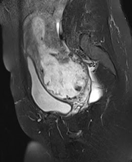
Sagittal T2FS image showing vaginal and uterine agenesis

Axial image showing normal left ovary

Axial image showing absence of vagina
17 year old patient presenting with primary amenorrhoea. MR reveals uterovaginal agenesis. Ovaries and kidneys were normally present.

Combined uterovaginal agenesis is the most common type of class I mullerian duct anomalies.
In Mayer-Rokitansky-Kuster-Hauser syndrome, patients usually have  agenesis or hypoplasia of upper two-thirds of vagina with intact ovaries. There may be variable anomalies of uterus, urinary tract and vertebrae.

Monday, 7 July 2014

MILWAUKEE SHOULDER

T2 GRE-AXIAL

T2FS-AXIAL PLANE


PDFS-CORONAL PLANE


STIR-SAG PLANE


H
T1-SAG PLANE

 60 year old female with shoulder pain and decreases range of motion: Above images show large globular foci of hypointense signal on all sequences in the infraspinatous tendon/ associated bursa  s/o calcification due to Hydroxyapatite deposition disease. There is fluid in adjacent bursa / oedema in adjacent soft tissues.

Thursday, 22 May 2014

TALOCALCANEAL COALITION

STIR coronal image

STIR coronal image
PDFS-sagittal image

T1-sagittal image

T1-sagittal image

Coronal and sagittal T1, STIR and PDFS sequences in a 30 year old female showing talocalcaneal coaltion.
There is continuity of marrow fat from the talus through the sustentaculum tali.
Secondary signs of talocalcaneal coalition are seen with talar beaking ( on last image).

This 30 year old female presented with rearfoot pain localized to the medial part of the foot along with flat foot.


Talocalcaneal coalition respresents 37 % cases of tarsal coalition with calcaneo-navicular coalition being the most comon type( 53 %).
Talocalcaneal coalition can occur through any of the 3 facets but is most common in the middle facet through the sustentacumum tali. These are best evaluated  on coronal images.
In fibrous coalition, there may be subchondral marrow oedema.

Secondary signs of a talocalcaneal coalition include:
Talar beaking adjacent to talonavicular articulation- also seen on lateral x-rays
Degenerative dorsal changes in calcaneocuboid joint

REFERENCES:
  • Stoller DW, Ferkel RD: The Ankle and Foot. Magnetic Resonance Imaging in Orthopaedics and Sports Medicine: Volume I: Third Edition. Lippincott Williams & Wilkins:1009-1013, 2007.
  • Radiopaedia.org


Friday, 28 February 2014

CERVICO-ISTHMIC PREGNANCY


A patient presented in her mid thirties with a first trimester intra-uterine pregnancy having conceived after IVF.  USG showed a fetus with cardiac activity and crown-rump length corresponding to 11 weeks' gestation.  The gestation sac was abnormally low lying with an empty endometrial cavity above it. The cervix was poorly visualized. An MRI was indicated to rule out true cervical pregnancy.

MRI showed a low lying gestation sac with empty endometrial cavity above it. The cervix was shortened with a closed cervical canal and internal os. There was a large low lying placenta surrounding the sac with loss of interface between the placenta and myometrium. The myometrium was thinned out. A diagnosis of cervico-isthmic pregnancy was made with the likelihood of adherent placenta.

CERVICO-ISTHMIC PREGNANCY


INTRODUCTION: A cervico-isthmic pregnancy is an ectopic pregnancy which implants in the isthmus. The isthmus is the region between the cervix and fundus.

It is a rare complication of pregnancy and is often confused with a cervical pregnancy. Distinguishing between these is crucial as a cervical pregnancy is virtually not viable while there are a few reports of cervico-isthmic pregnancies being carried successfully to term. Also, cervical pregnancies are more dangerous and associated with serious risks like haemorrhage, rupture, blood transfusions, hysterectomy etc.

INCIDENCE:
Incidence of cervical pregnancy: 1:1000 to 1:16000.
Cervico-isthmic pregnancies are more common than true cervical pregnancies.

IMAGING FINDINGS:
USG: Typically, ultrasound shows an empty uterine cavity with a low lying pregnancy. It may be confused with true cervical pregnancy. In a true cervical pregnancy, the sac is completely within the cervix.

MRI: shows a  low lying gestation sac with a well preserved and closed cervical canal. It is hence helpful in distinguishing cervical from cervico-isthmic pregnancy.

IMPLICATIONS: Although there are reports of successful cervico-isthmic pregnancies, these pregnacies are also exposed to risks like:
  • Spontaneous abortion/ premature delivery
  • premature rupture of membranes
  • placenta accreta
  • postpartum hemorrhage
  • hysterectomy
  • Uterine rupture if associated with thinned out myometrium
REFERENCES:
  1. Oyelese Y, Elliott T B, Asomani N, Hamm R, Napoli L, Lewis KM: Sonography and Magnetic Resonance Imaging in the Diagnosis of Cervico-Isthmic pregnancy. J Ultrasound Med 2003; 22: 981-983
  2. Strobelt N, Locatelli A, Ratti M, Ghidini A: Cervico-isthmic pregnancy: a case report, critical appraisal of the diagnostic criteria, and reassessment of the outcome. Acta Obstet Gynecol Scand 2001; 80: 586-588.
  3. Jelsema RD, Zuidema L: First trimester diagnosed cervico-isthmic pregnancy resulting in term delivery. Obstet Gynecol 1992; 80: 517-519
  4. Wikipedia


RHOMBENCEPHALOSYNAPSIS

Figure 1: Coronal T2 image showing fused dentate nucleus
Figure 2: Coronal T2 weighted image showing typical transverse folia and sulci with fused cerebellar hemispheres

Figure 3: Axial T2WI showing fusion of the interfoliate sulci across midline along with fusion of cerebellar folia.

Figure 4: Axial T2WI showing "keyhole appearanca" of 4th ventricle and midline fusion of cerebellar white matter. Vermis is not seen.

Figure 5: Sagittal T2WI showIng absent primary fissure. There is also slight rounding of the fastgial recess. Note the corpus callosum in a patient of isolated rhombencephalosynapsis.

The above MR was done in an adult patient presenting with mild gait ataxia. Rhombencephalosynapsis was detected in him as an isolated finding.

  • Rhombencephalosynapsis is a rare  posterior fossa malformation characterized by aplastic/ hypoplastic cerbellar vermis and fusion of cerebellar hemispheres in the midline.
  • It was forst described by Obersteiner in 1914.
  • Pathogenesis is related to disturbed cerebellar development between 28 to 41 days of gestation.
  • Clinical profile varies and is related to the posterior fossa malformation and associated supratentorial anomalies. It might vary from mild truncal ataxia and normal intelligence to cerebral palsy.
  • Isolated rhombencephalosynapsis is less common than rhombencephalosynapsis associated with supratentorial anomalies
  • Most cases are reported in infancy or pediatric age group with only few cases reported in adults.

Diagnosis is usually by MRI which is clearly superior to CT in evaluating these patients.

Distinctive radiological features are:
Posterior fossa:
  • Agenesis / hypoplastic vermis 
  • Absent posterior cerebellar incisura, vallecula
  • agenesis or hypogenesis of anterior vermis ( rostral ) with hypoplastic posterior vermis( caudal  nodulus may form
  • Midline fusion of cerebellar hemispheres to form a single lobed cerebellum
  • Fissures and cerebellar folia are transversely oriented best seen on coronal MRI
  • Superior and middle cerebellar peduncles may be fused along with dentate nuclei giving a characeteristic diamond shaped, posteriorly pointing 4th ventricle
Supratentorial anomalies:
  • Most common supratentorial anomaly is hydrocephalus with aqueductal stenosis
  • Sometimes, there may be fused thalami, fornIces, cerebral peduncles, inferior collicluli with absent septum pellucidum
  • Dysgenetic limbic system
  • cortical malformatins
  • sutural synostosis
  • hypoplastic commissures, hypoplastic anterior visual pathway and agenesis of posterior lobe of pituitary gland
  • Association with septo-optic dysplasia and holoprosencephaly
 References:
  • Mendonca JLF, Natal MRCR, Viana SL et al. Rhombencephalosynapsis: CT and MRI findings. Neurology India;152:118-120, 2004.
  • Weerakkody Y, Gaillard F et al. Rhombencephalosynapsis. Radiopaedia.org.
  • Blaser SI: Rhombencephalosynapsis. Diagnostic Imaging Brain: First Edition. Amirsys: I-1-30, 2005.

Wednesday, 29 January 2014

IDIOPATHIC INTRACRANIAL HYPERTENSION

IDIOPATHIC INTRACRANIAL HYPERTENSION:


 Figure 1: showing increased fluid in sheaths surrounding optic nerves along with tortuosity of the sheaths
 Figure 2: showing flattening of posterior sclera
Figure 3: Sagittal T2 WI showing a partially empty sella

The above pictures are of a 35 year old female who presented with headache and progressive visual loss.

MRI of the brain and orbits showed a partially empty sella, dilated/ tortuous optic nerve sheaths along with posterior scleral flattening. 
There was no evidence of an intracranial mass/ space occupying lesion or dural sinus thrombosis.

IDIOPATHIC INTRACRANIAL HYPERTENSION( IIH):
Also called pseudotumor cerebri.
As the name implies,the intracranial pressure is raised with no obvious underlying pathology in the brain.
Most common clinical presentation is an obese female aged 20-40 years with headache( aggravated by Valsalva), papilledema, progressive visual loss, diplopia etc.
MRI reveals a partially empty sella, optic nerve sheath dilatation with vertical tortuosity, optic nerve head protrusion and scleral flattening.
The diagnosis is made after other potential causes of raised intracranial hypertension like dural sinus thrombosis, space occupying lesion are excluded.
Aim of treatment is to prevent visual loss which is a potential danger from chronic papilledema.

REFERENCES:
  1.  Suzuki H, Takanashi J, Kobayashi K et al: MR Imaging of Idiopathic Intracranial Hypertension. AJNR 22:196-199, 2011
  2. Hingwala DR, Kesavadas C, Thomas B et al: Imaging signs in idiopathic intracranial hypertension: Are these signs seen in secondary intracranial hypertension too? Ann Indian Acad Neurol 16(2):229-233, 2013
  3. Castillo M: Idiopathic Intracranial Hypertension. Diagnostic Imaging Brain: First Edition. Amirsys: I-10-36, 2005.